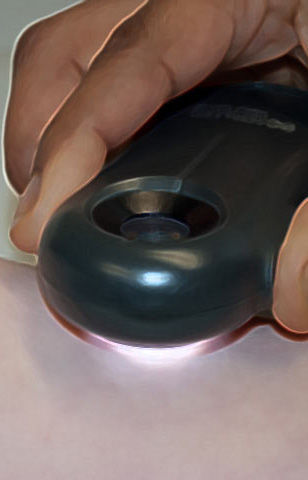
Virtual Dermatoscope
 A flat pigmented lesion on a 20 year old man’s back
 A palpable pigmented lesion on a young girl's thigh
 A pigmented lesion on a 10 year old girl's index finger
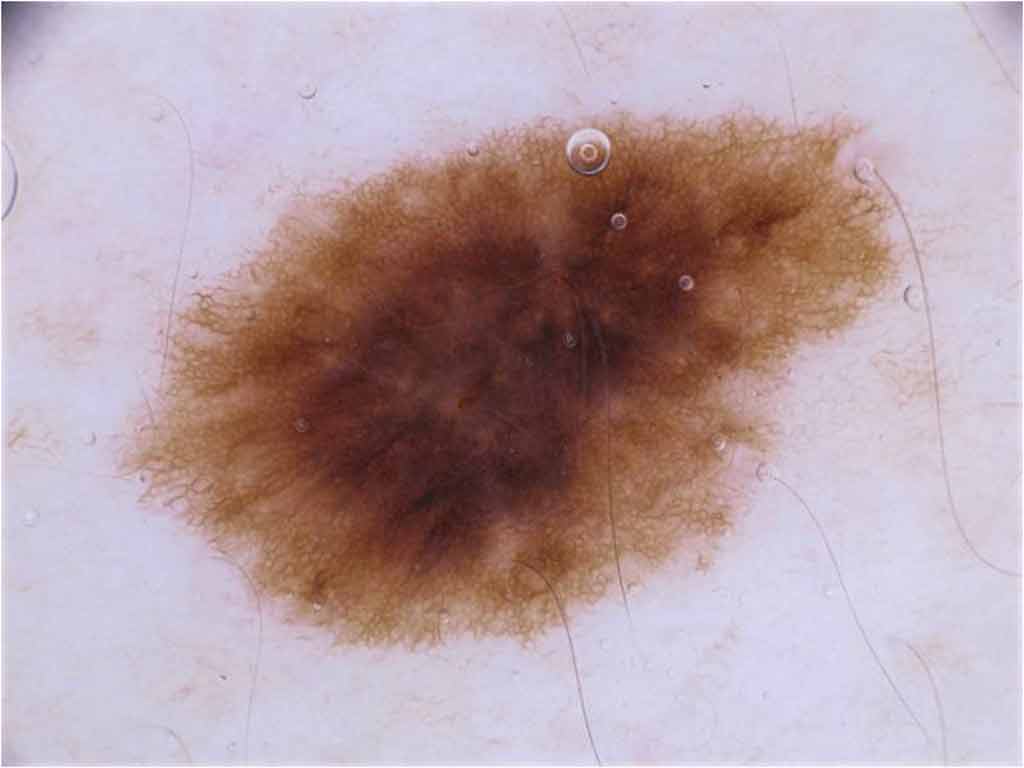
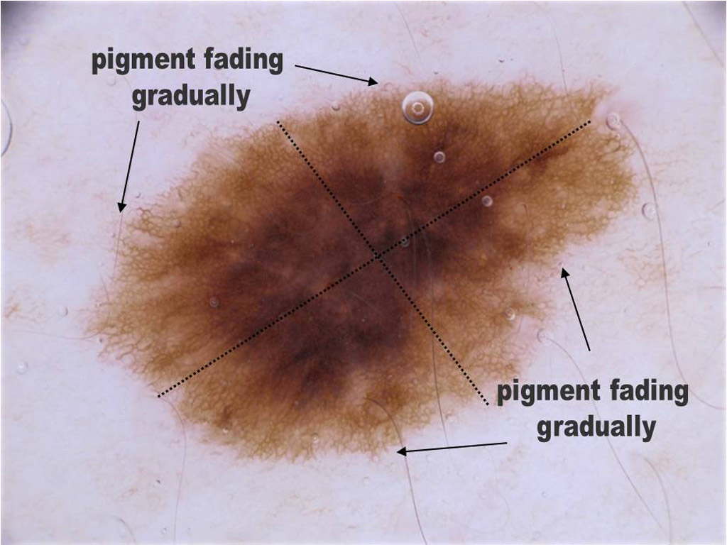
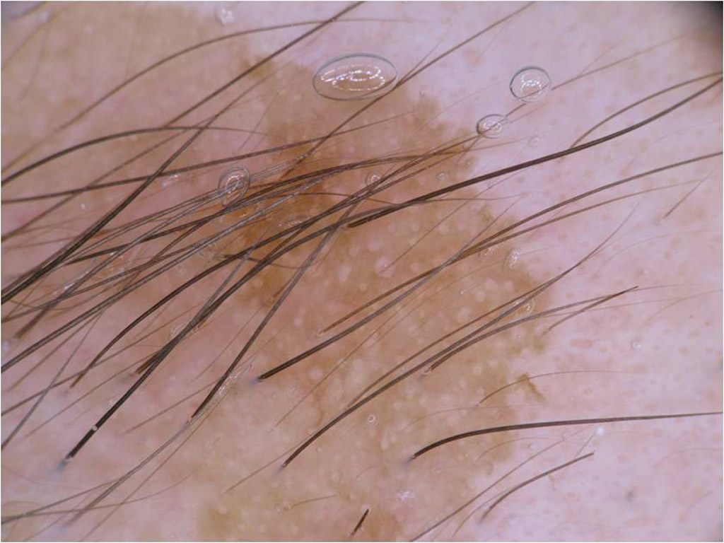
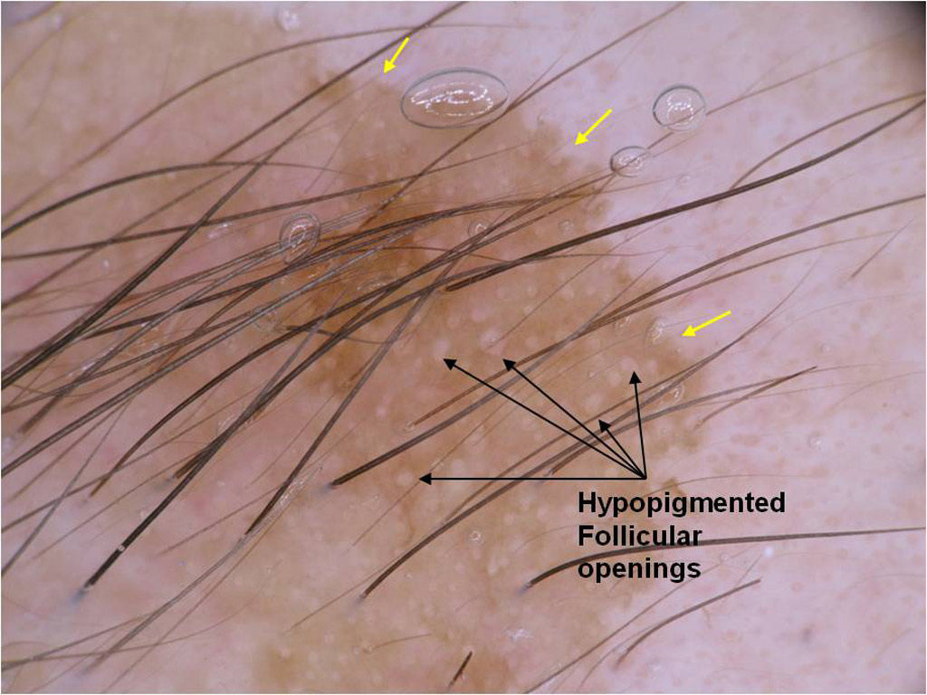
 This lesion on a 40 year old woman's leg has not changed in 15 years
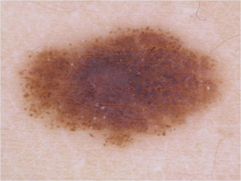
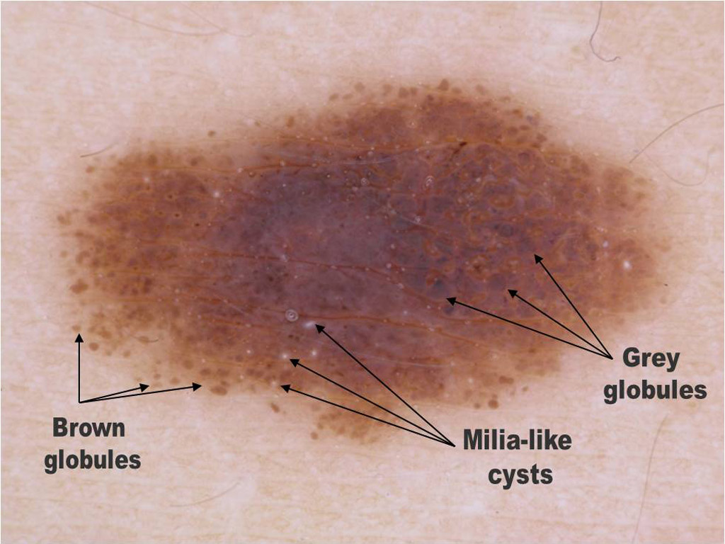
 A palpable pigmented lesion on a 58 year old man's back
 A palpable lesion on a 38 year old man’s abdomen
 A flat pigmented lesion on the areola
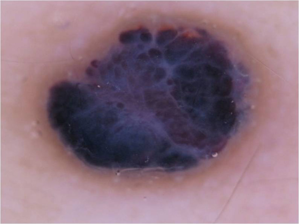
 Rapidly growing heavily pigmented nodule. 32 year old man's abdomen
 Large pigmented macule on face of a woman with sun-damaged skin