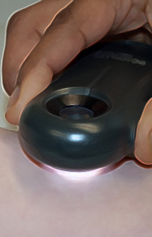
Virtual Dermatoscope
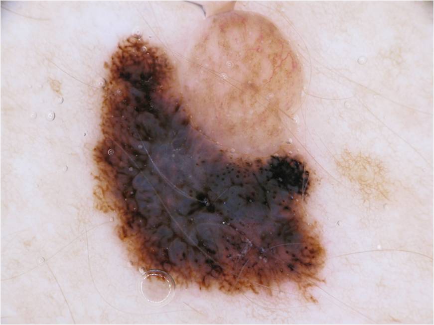
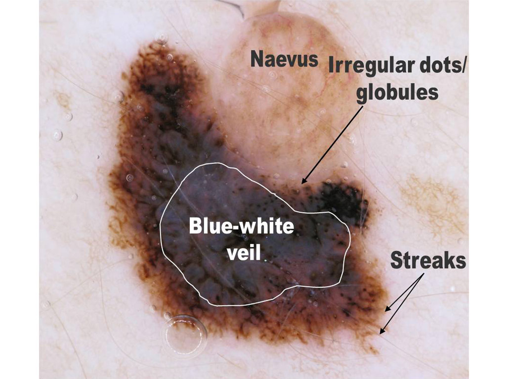
 A palpable pigmented lesion on the back of a 60 year old woman
 A palpable lesion on the leg of a 58 year old woman
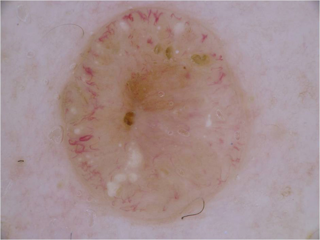
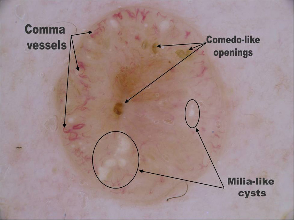
 A palpable lesion on a 47 year old man’s abdomen
 This is a pigmented lesion on a young man’s leg
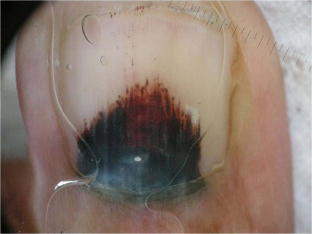
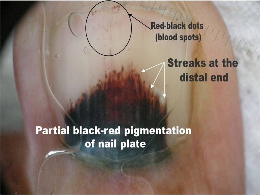
 Pigmentation of a fingernail of recent onset
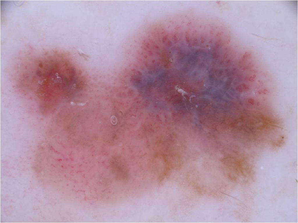
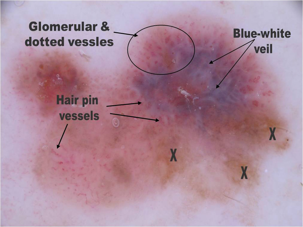
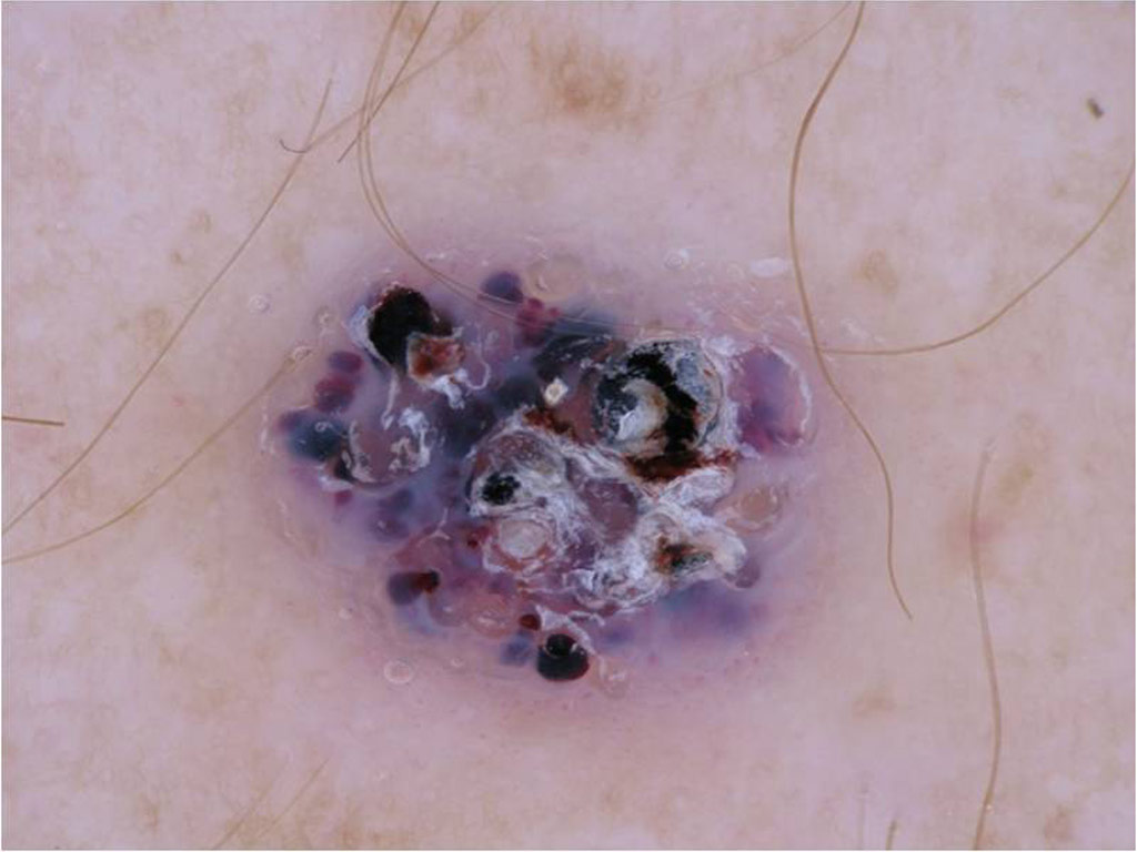
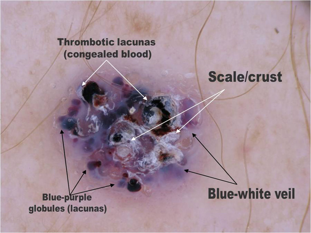
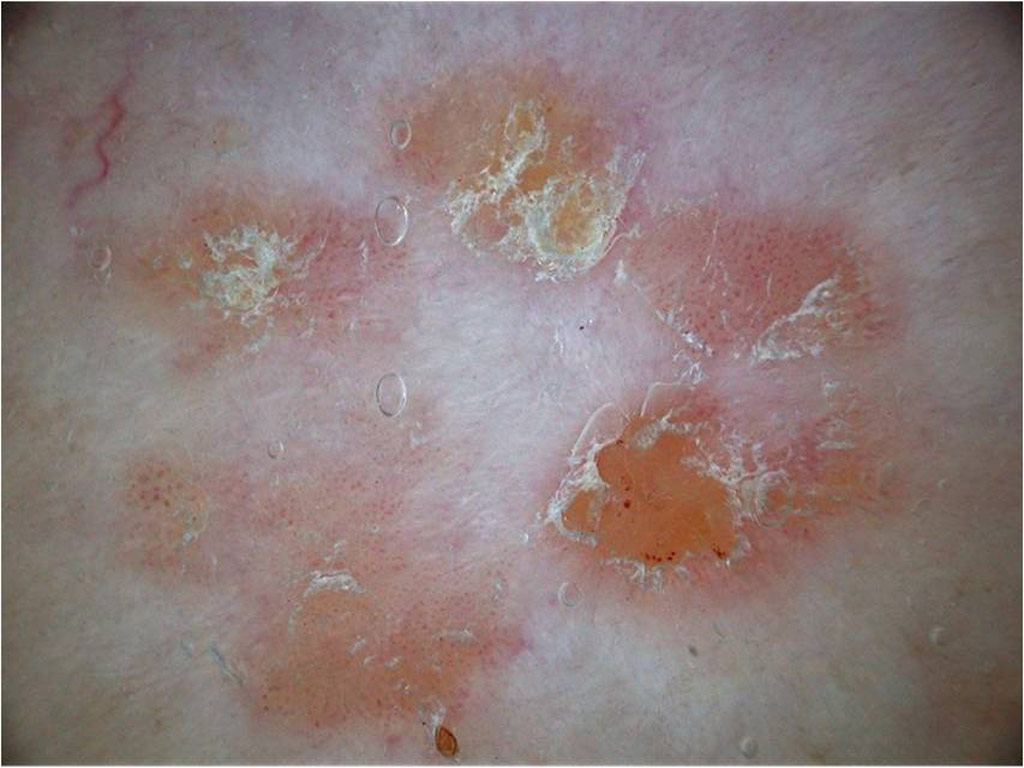
 An erythematous lesion on a 67 year old woman’s leg
 A pigmented lesion on the chest of a 40 year old man
 A small brown papule on the face of a young man
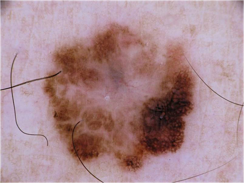
 A slightly pigmented lesion on a 42 year old lady’s leg