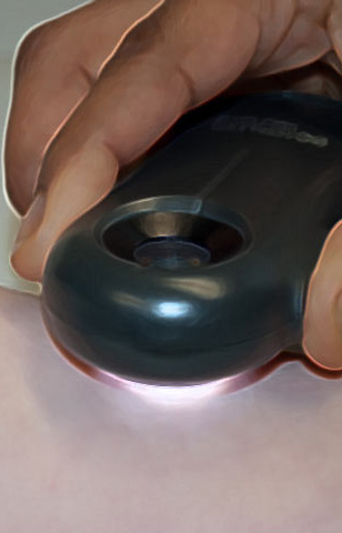
Virtual Dermatoscope
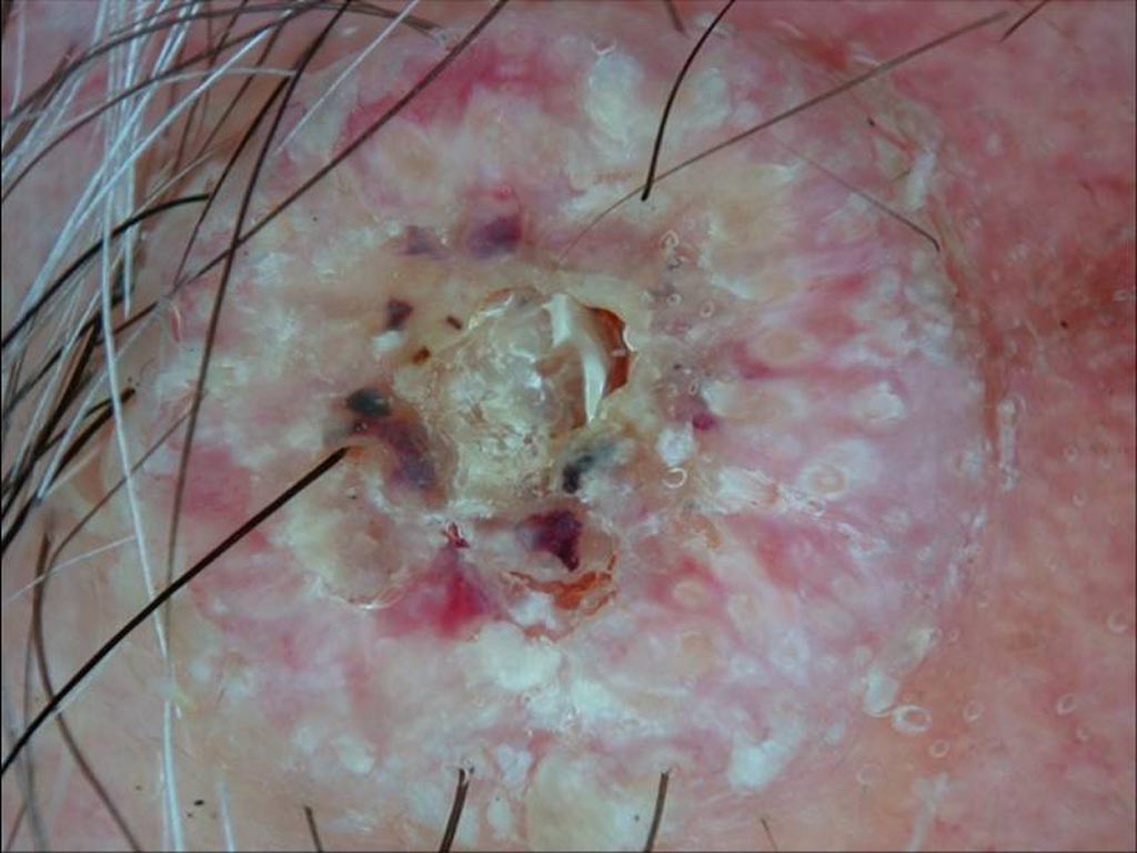
 Ulcerated lesion on right postauricular region of a 73 year old man
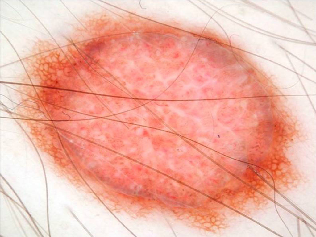
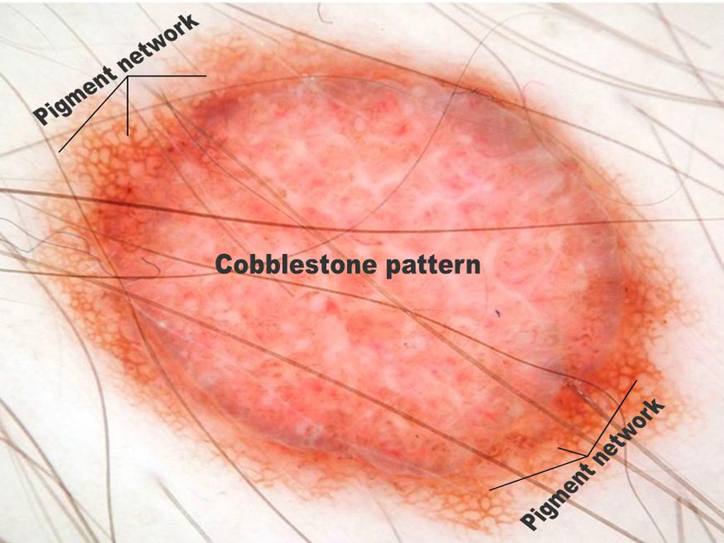
 Palpable lesion on a 35 year old man’s chest
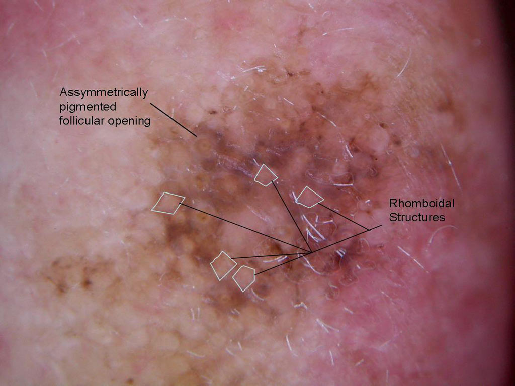
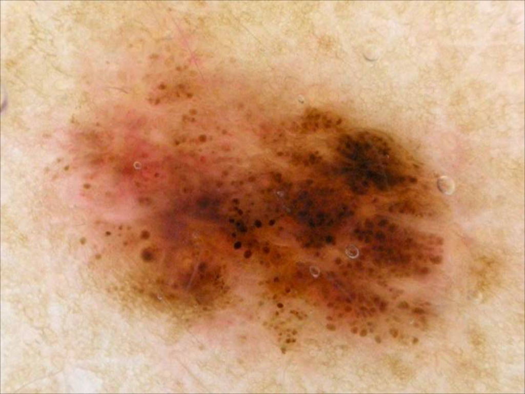
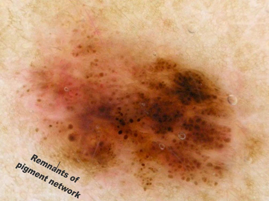
 A flat pigmented lesion on a 73 year old man’s ear
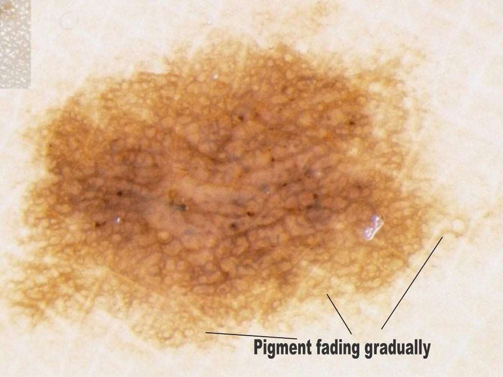
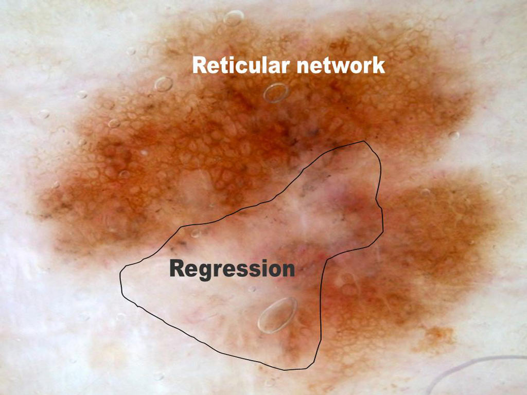
 A pigmented lesion on the trunk of a 59 year old man
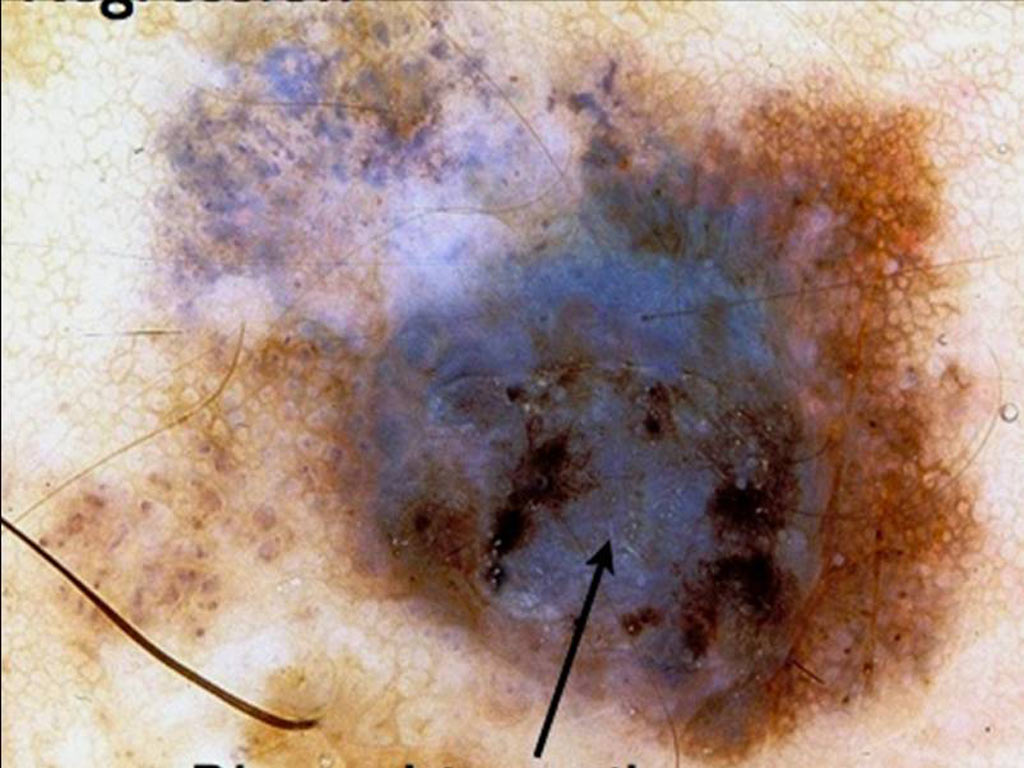
 A pigmented lesion on a young woman’s arm
 A palpable pigmented lesion on a 68 year old woman’s leg
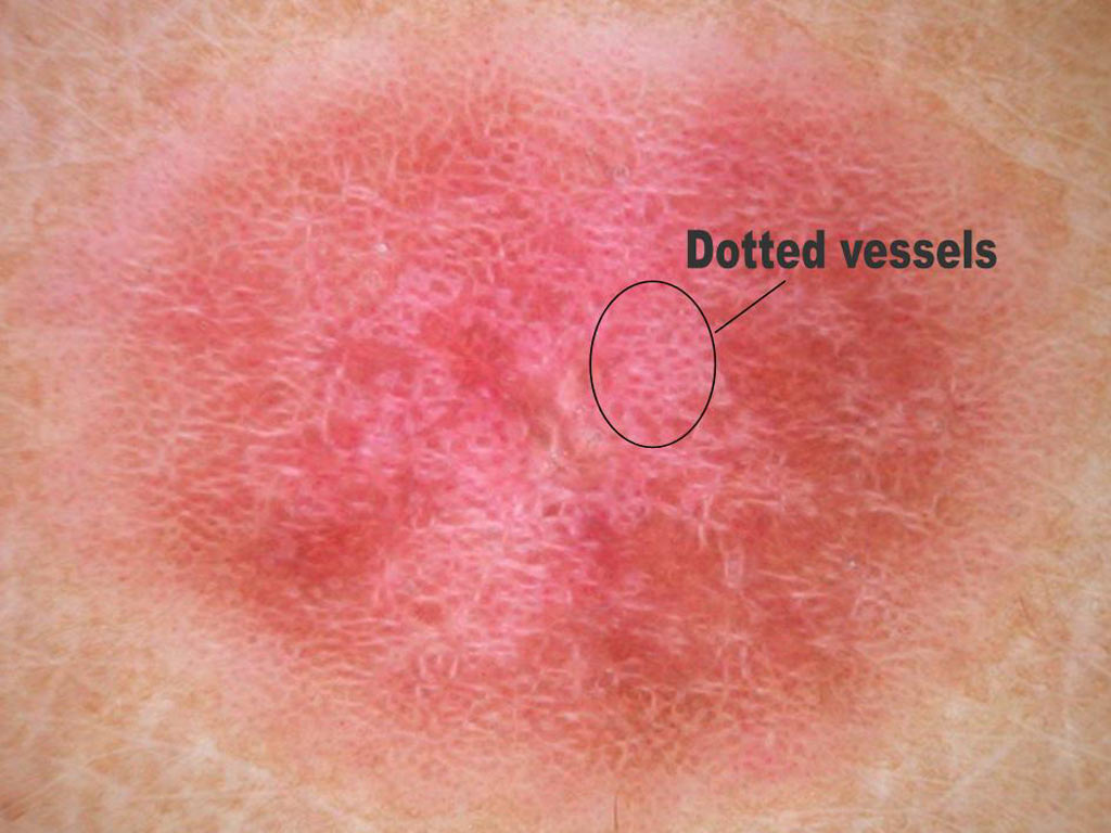
 An ill defined erythematous scaly lesion on a the forehead
 A pigmented lesion on a 36 year old man’s chest
 An erythematous lesion on a young girl’s thigh