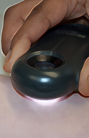
Virtual Dermatoscope
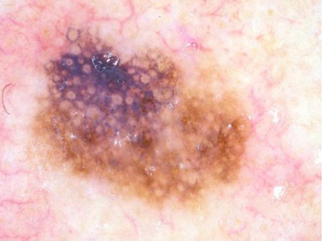
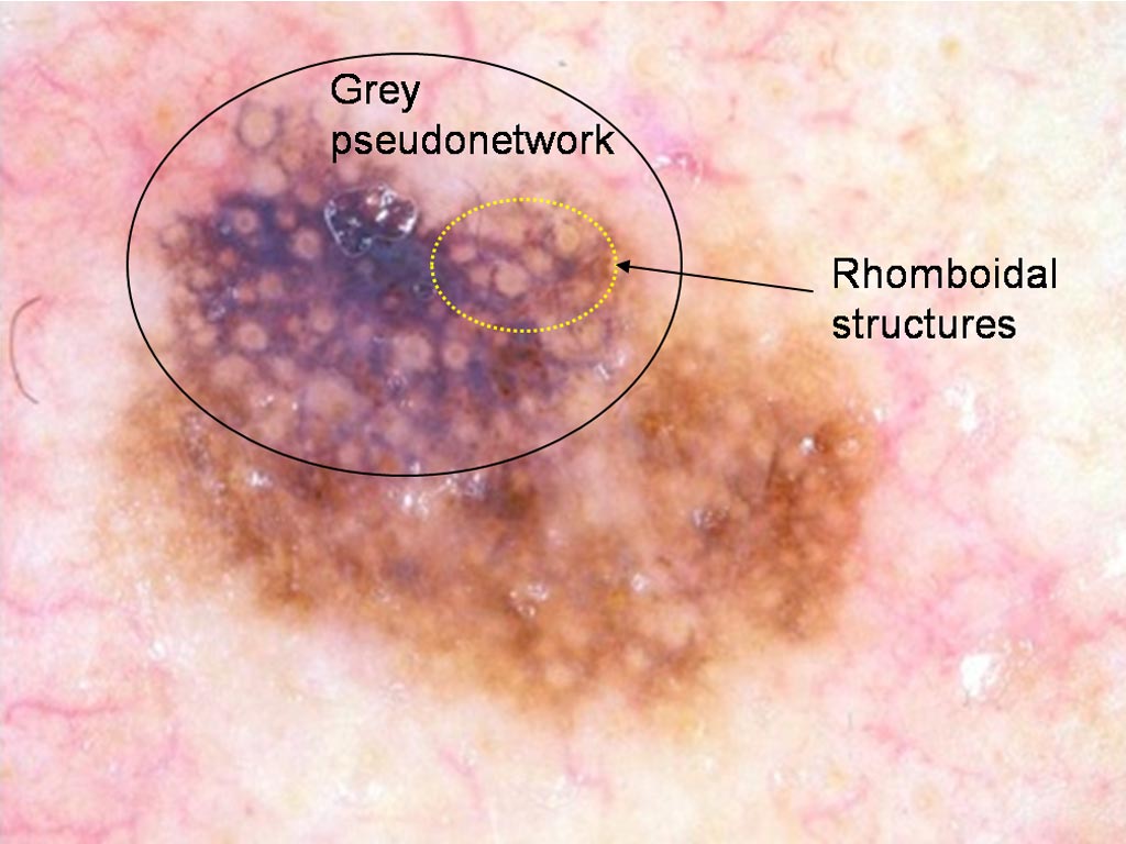
 Brown lesion on a 66 year old woman’s forehead
 Pigmented lesion on the foot of a 58 year old man
 Lesion on the face of an 80 year old man
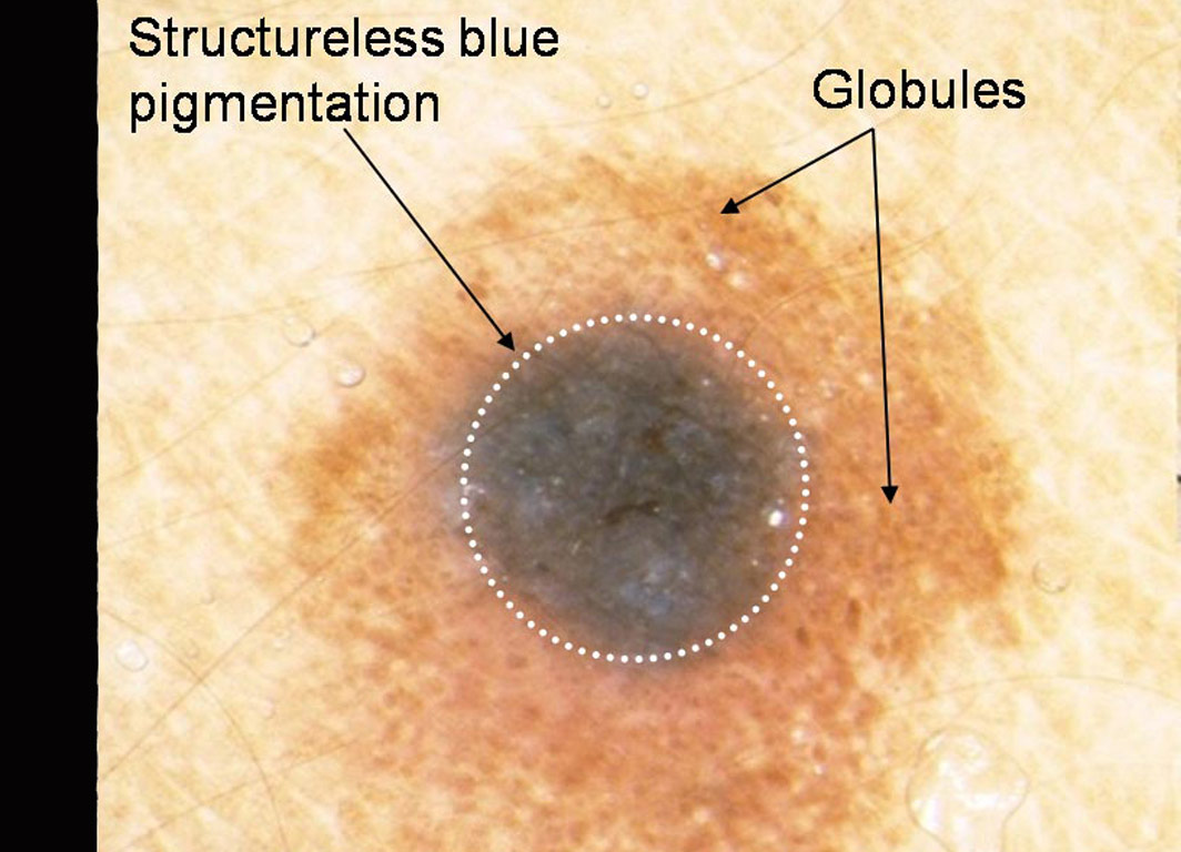
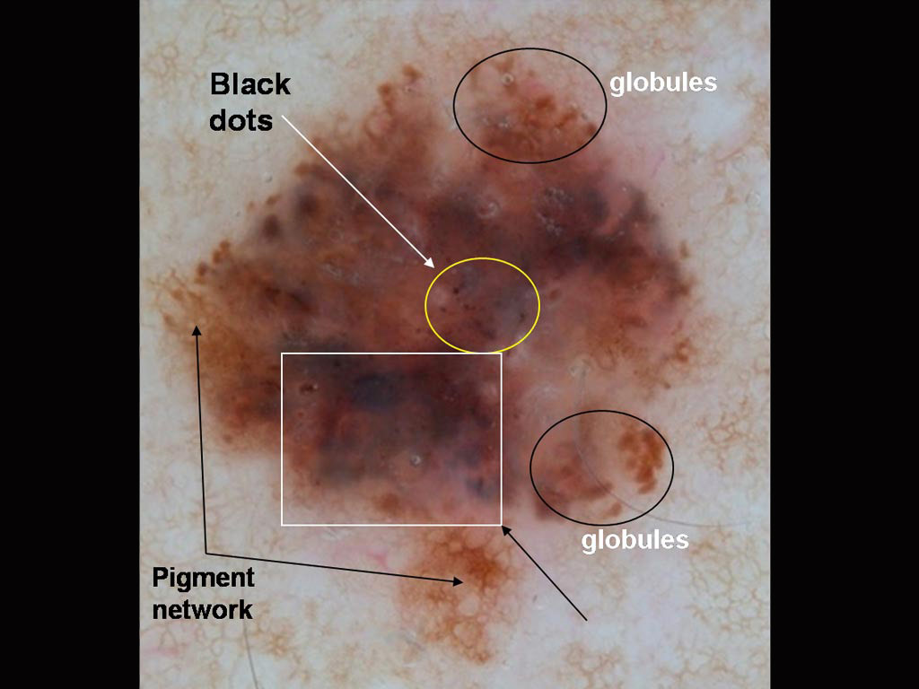
 A lesion on a 58 year old woman’s thigh
 A pigmented lesion on a 36 year old man’s chest
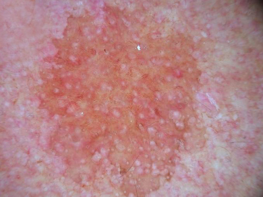
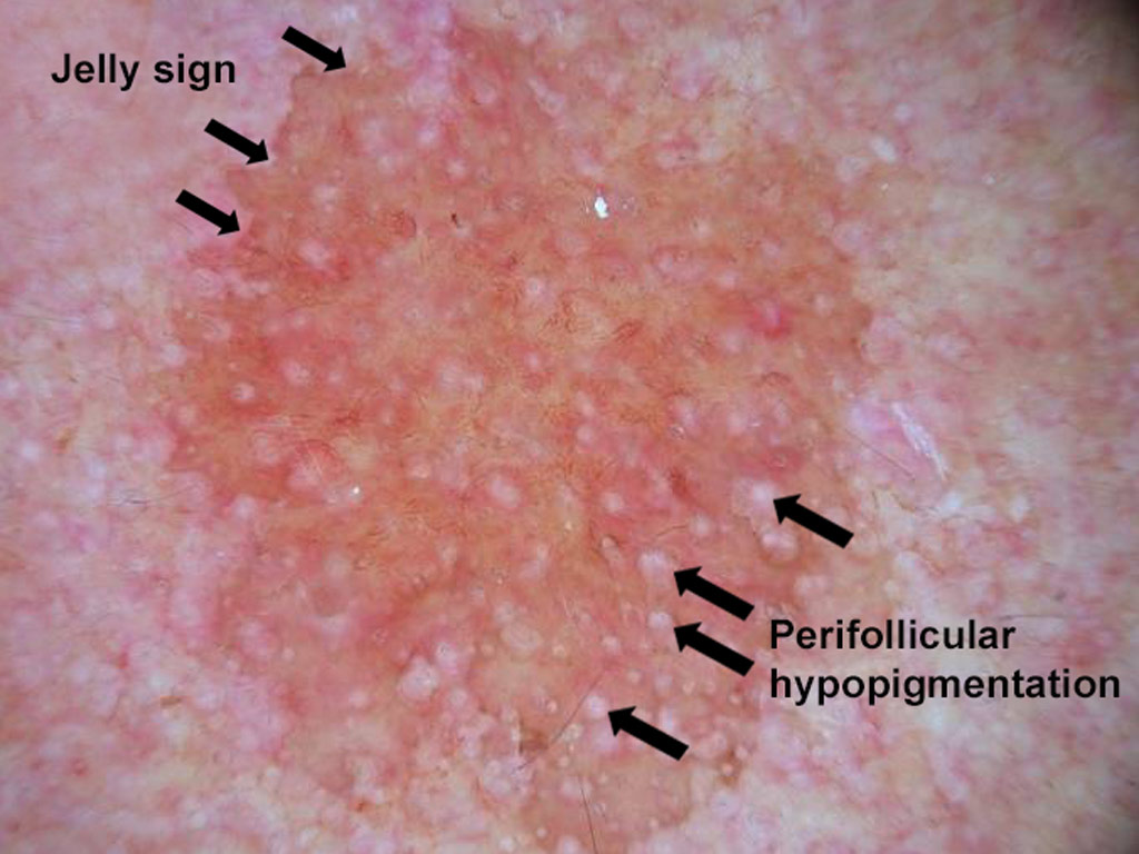
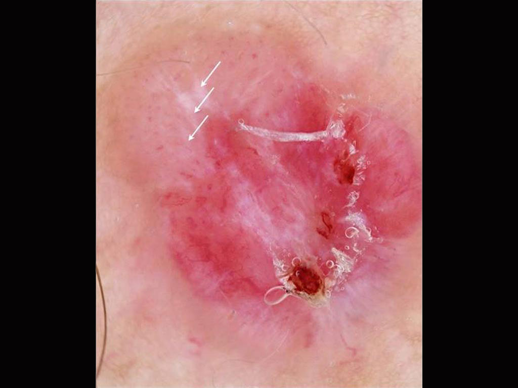
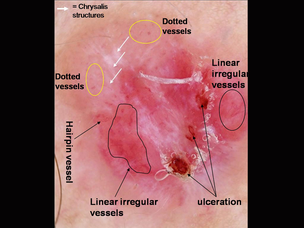
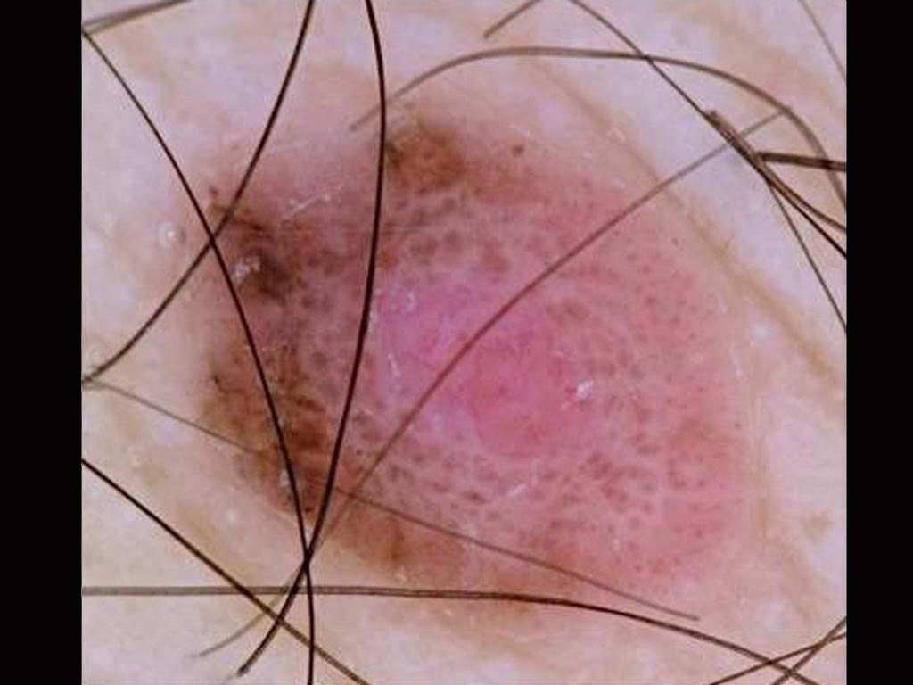
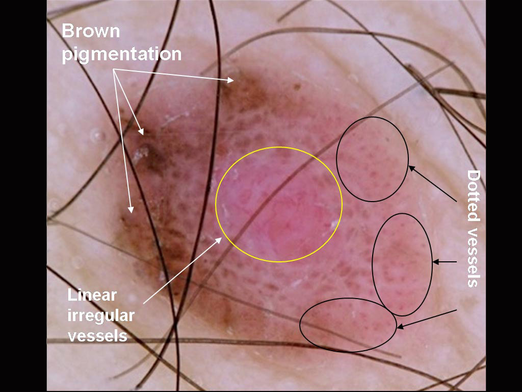
 A pink nodule on a young man’s back
 A pigmented lesion on an elderly woman’s abdomen
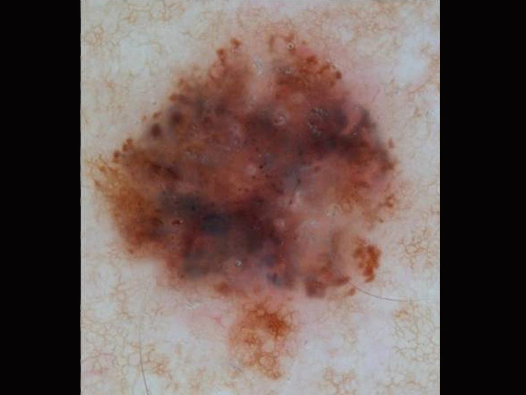
 Small pink lesion on a 47 year old man’s leg
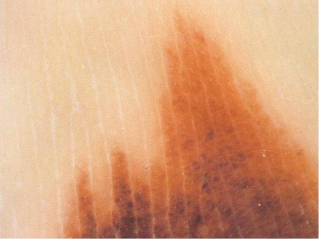
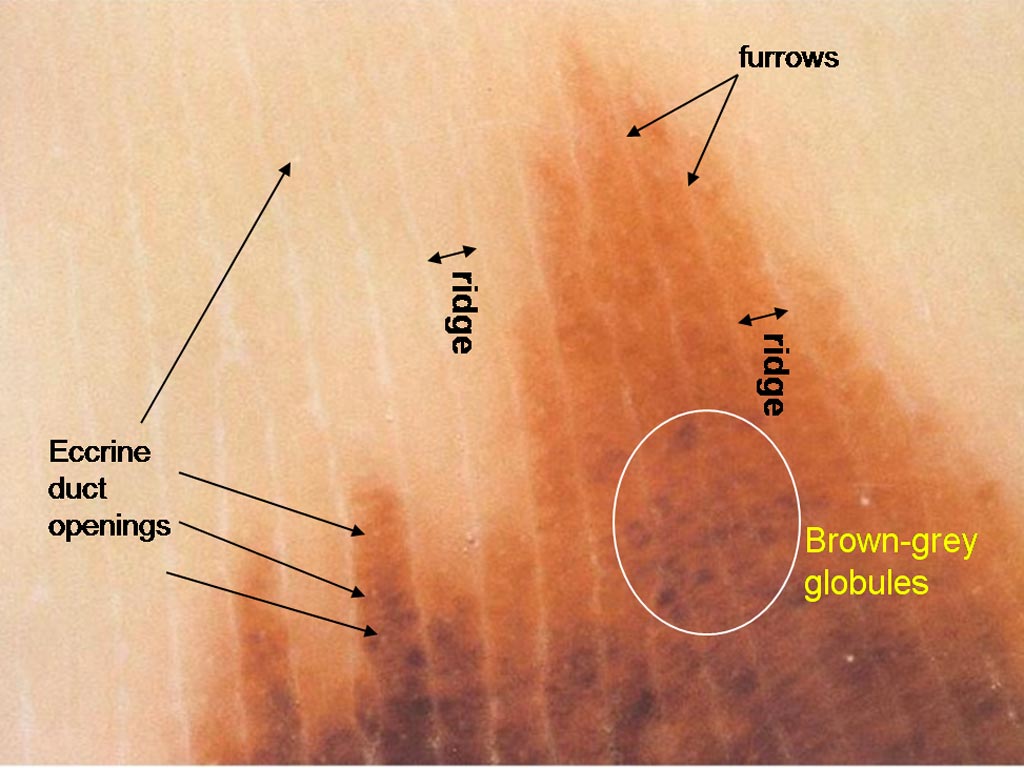
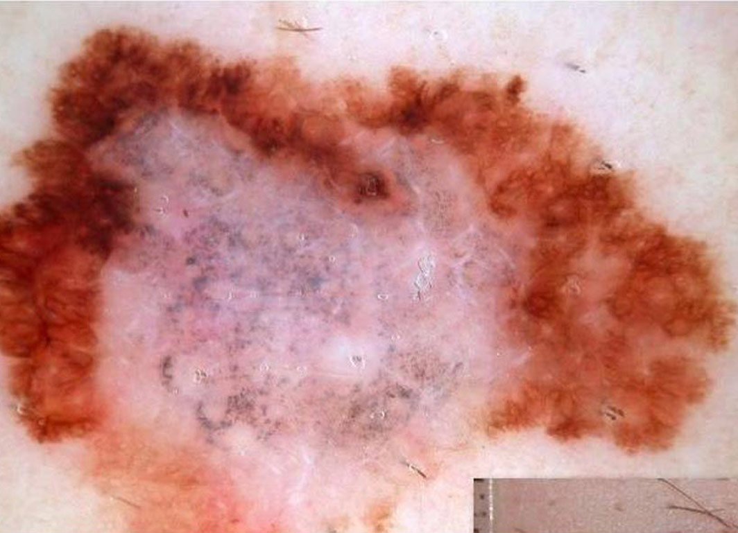
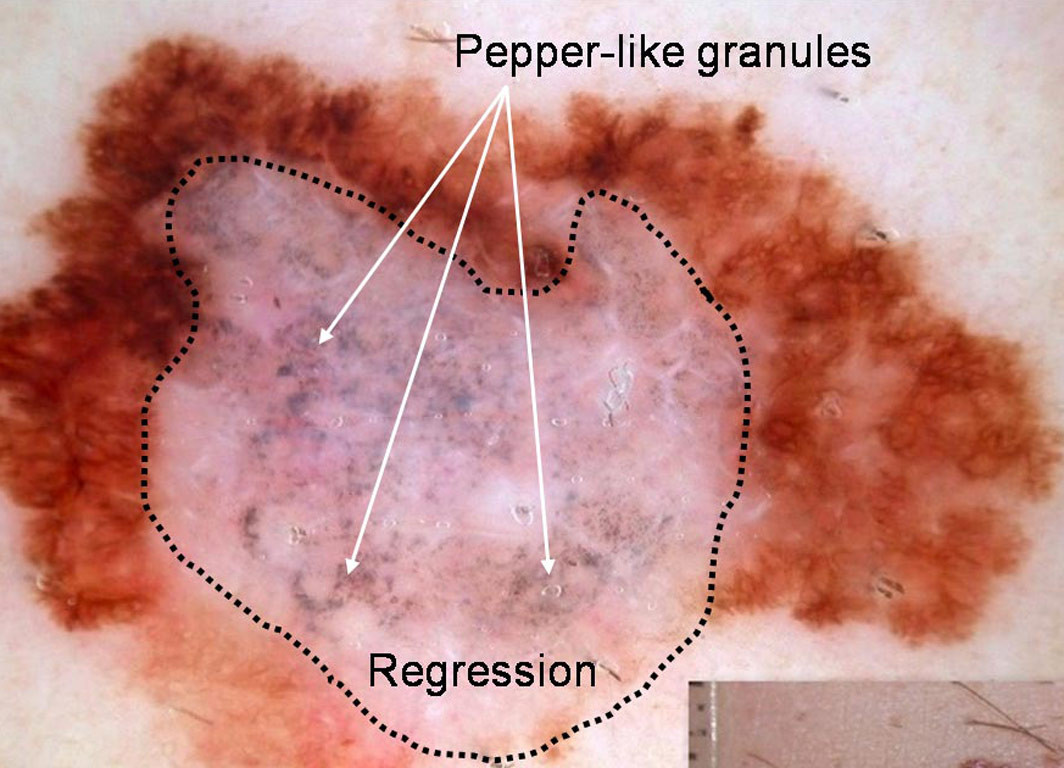
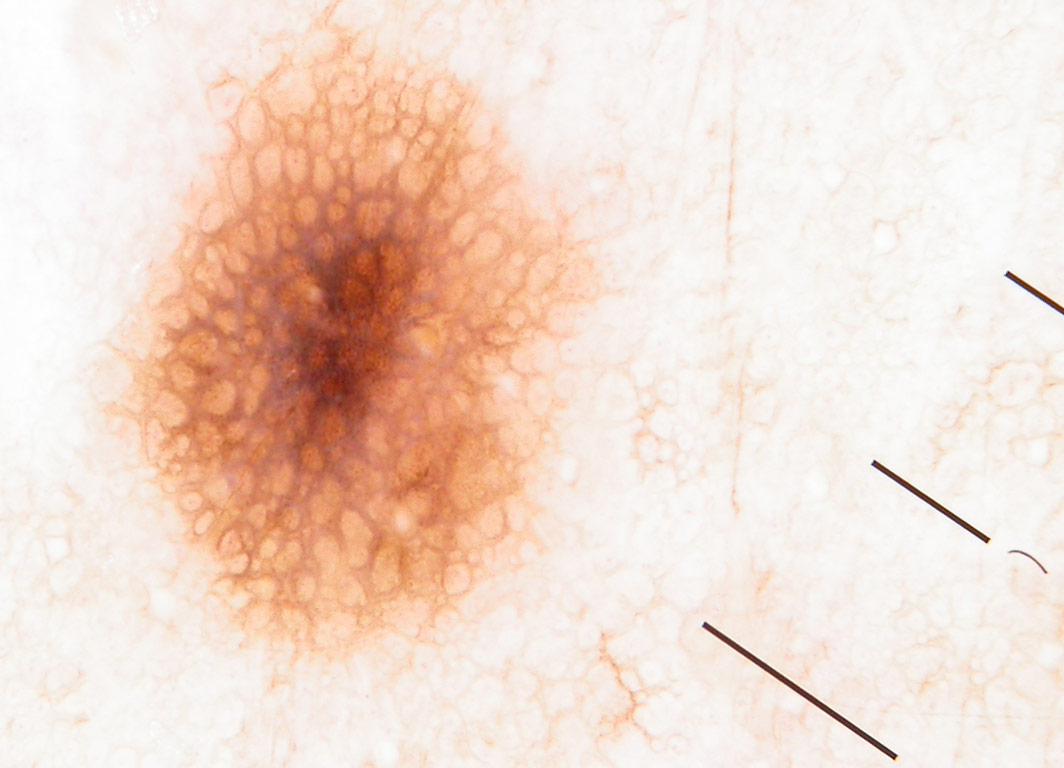
 A 50 year old man with fair skin has a flat pigmented skin lesion on his abdomen