Virtual Dermatoscope
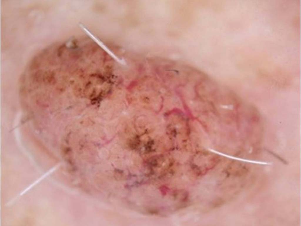
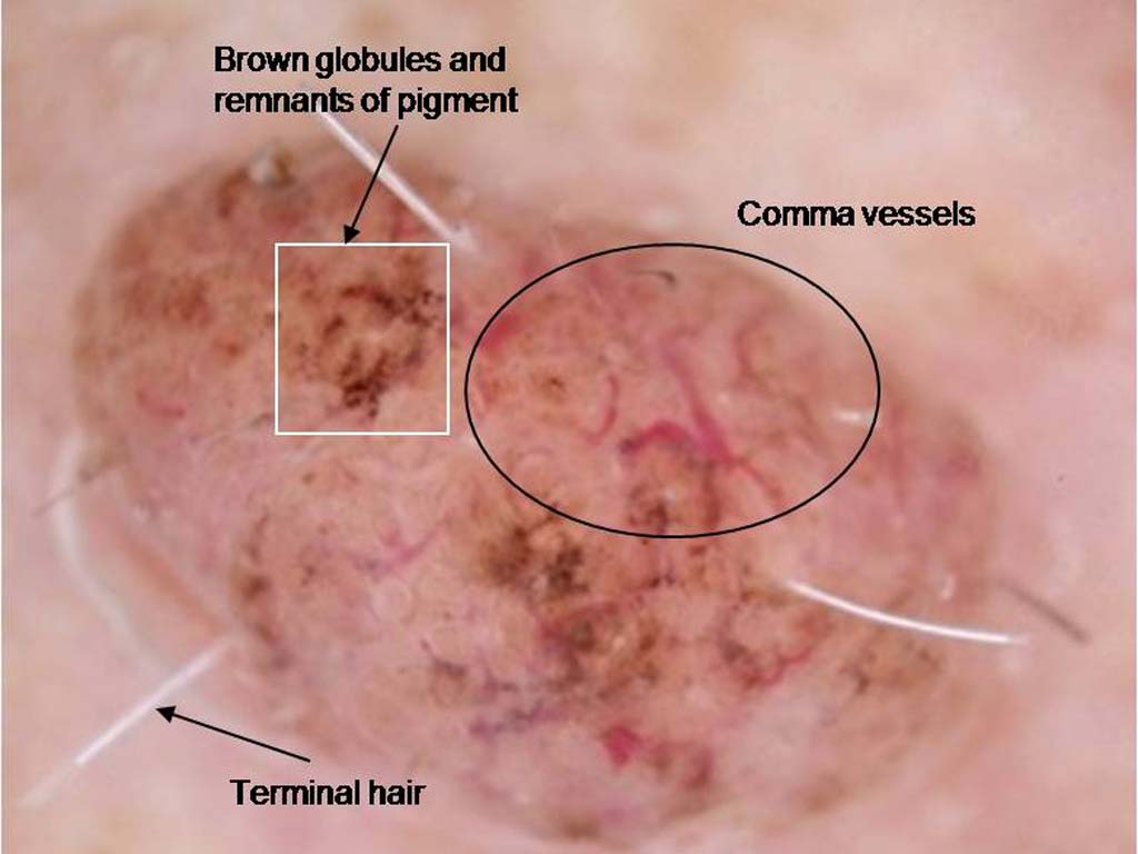
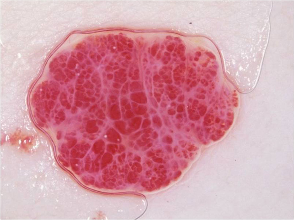
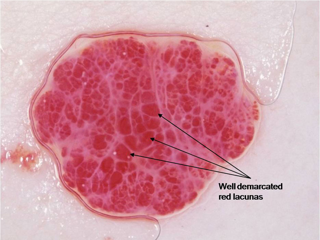
 This is a lesion on a 50 year old woman’s face
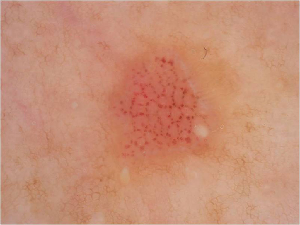
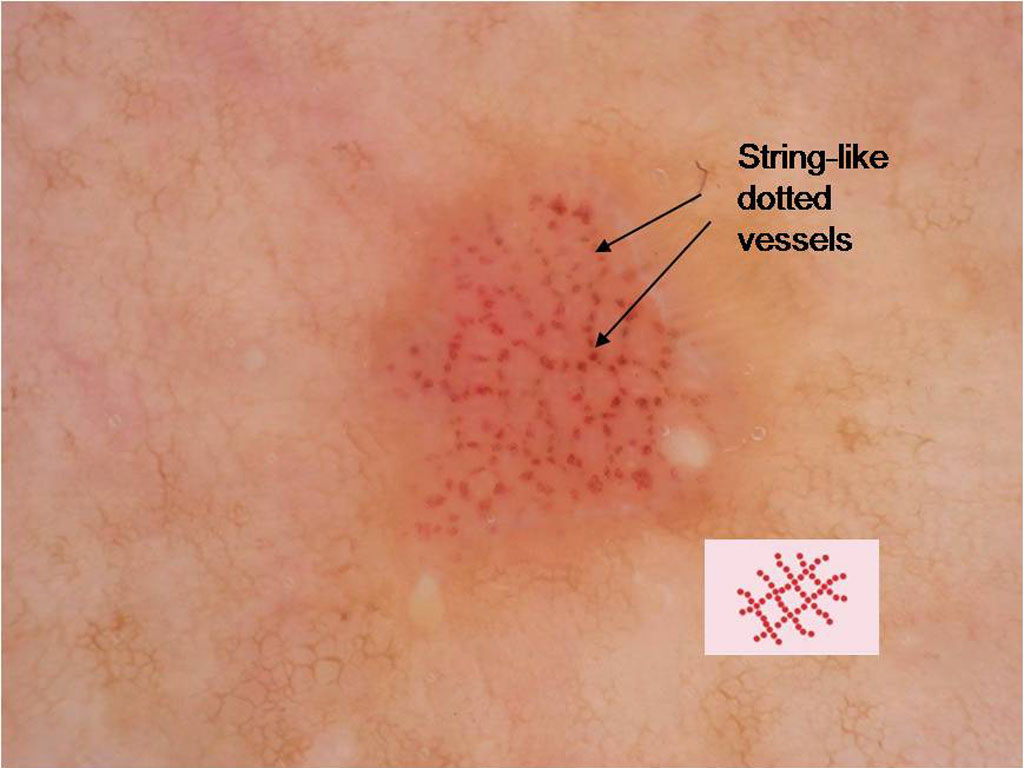
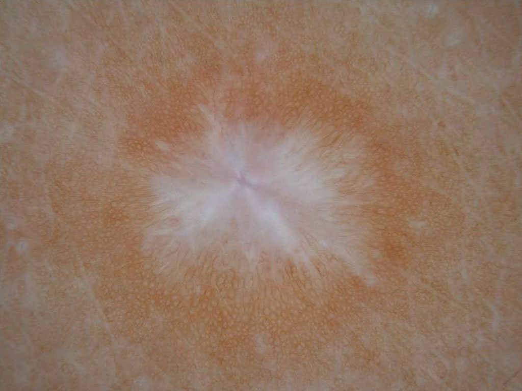
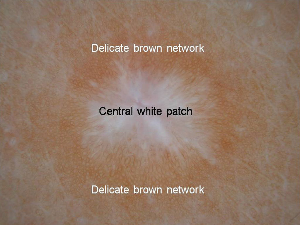
 This is a lesion on the chest of a 52 year old man
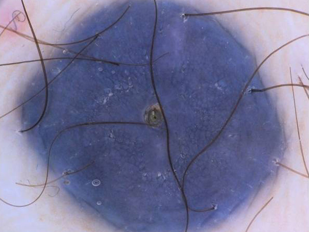
 This is a lesion on a chest of a 57 year old man
 This is a lesion on a 20 year old man’s leg
 This lesion has present for over 25 years with no change
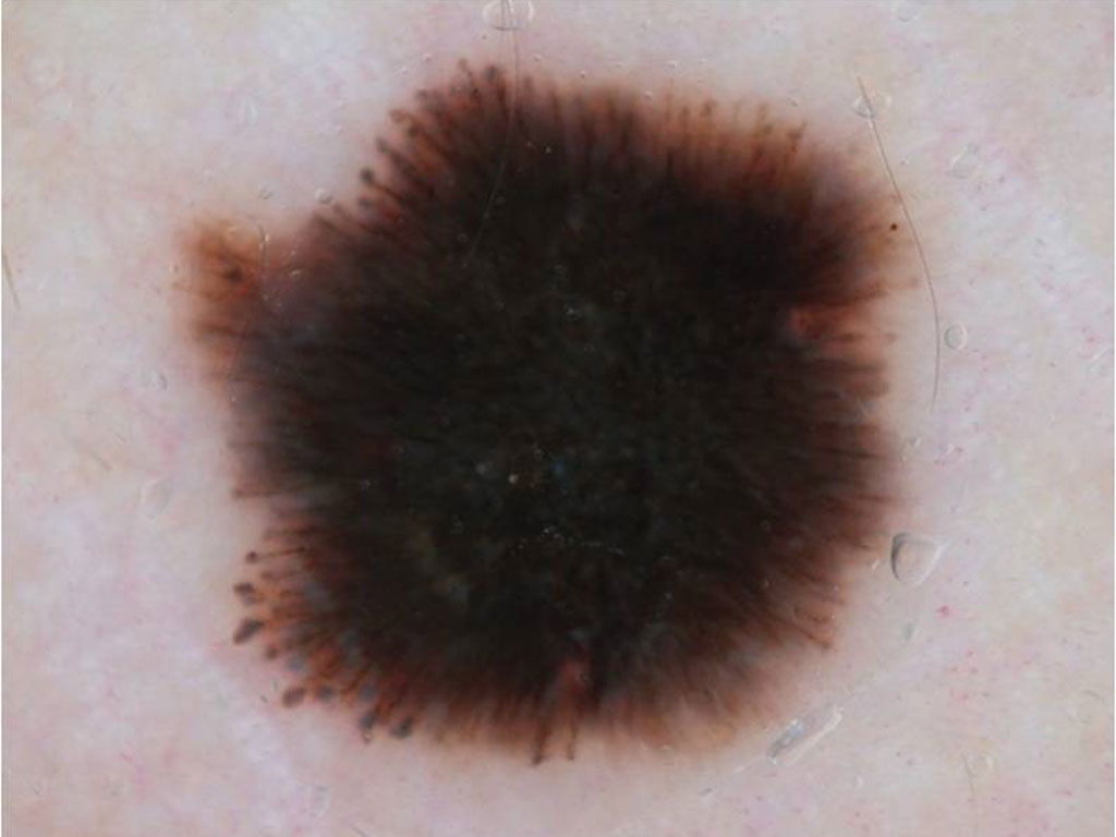
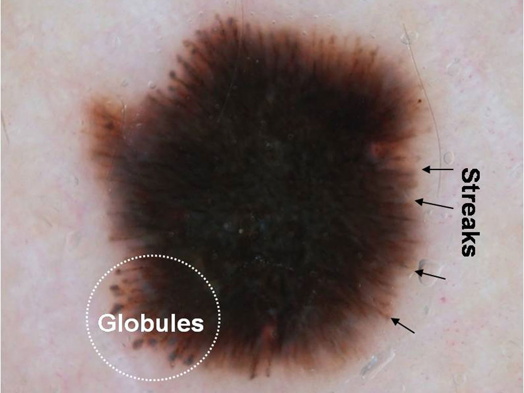
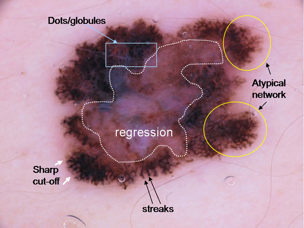
 Pigmented lesion on the back of a 12 year old boy
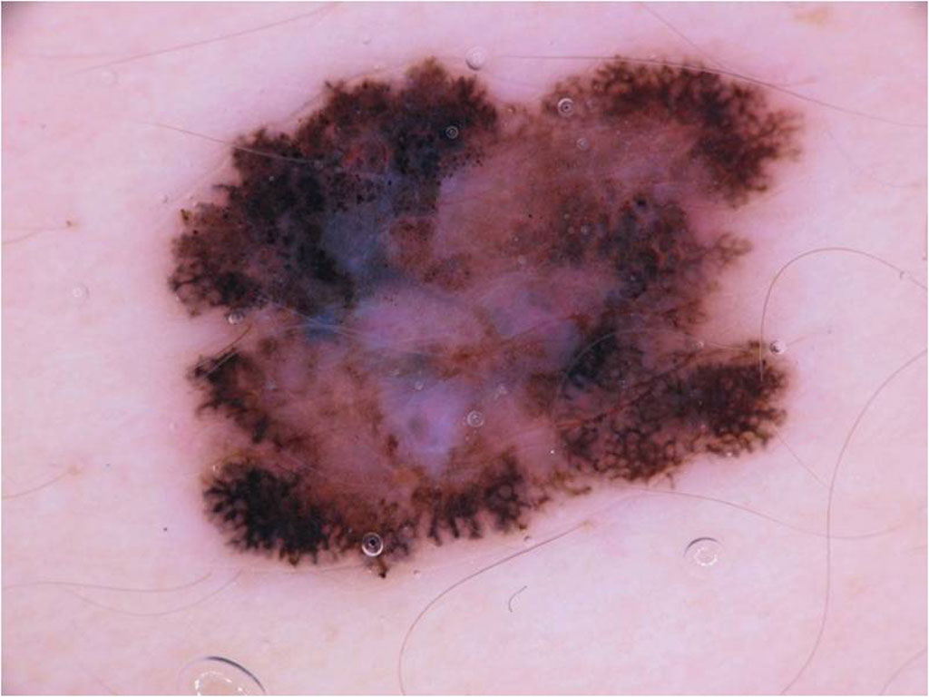
 Pigmented lesion on a 40 year old woman’s leg
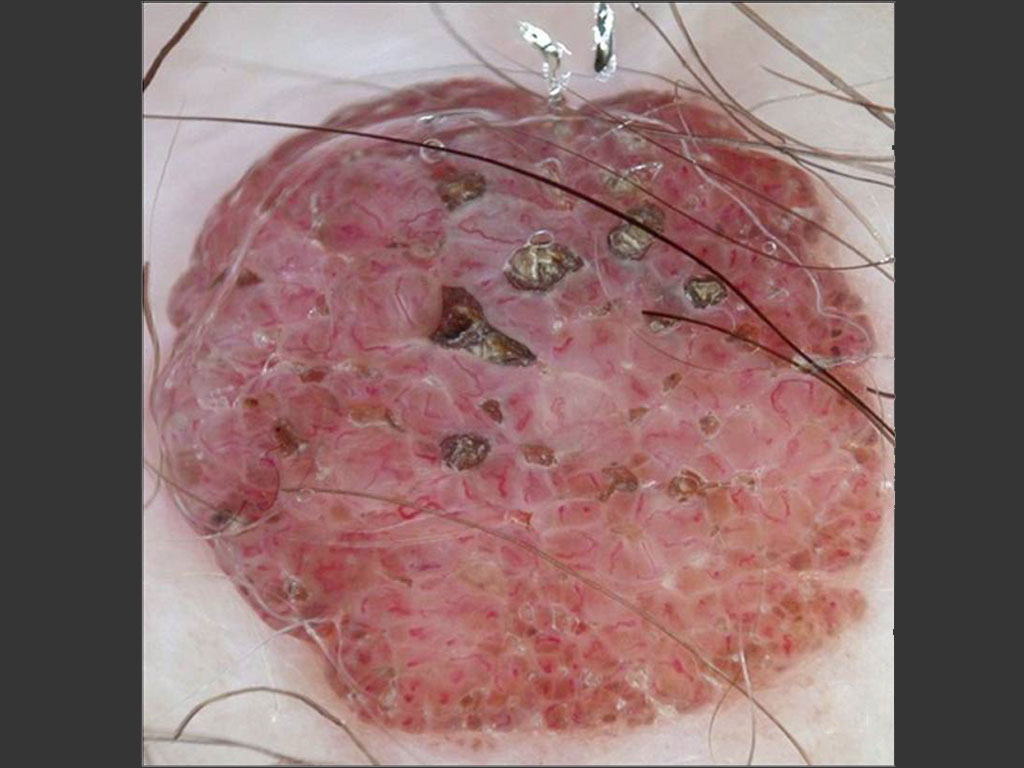
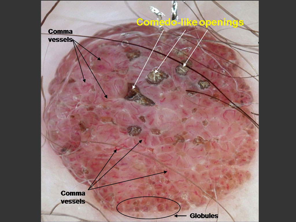
 A papillomatous lesion on a 54 year old man’s chest
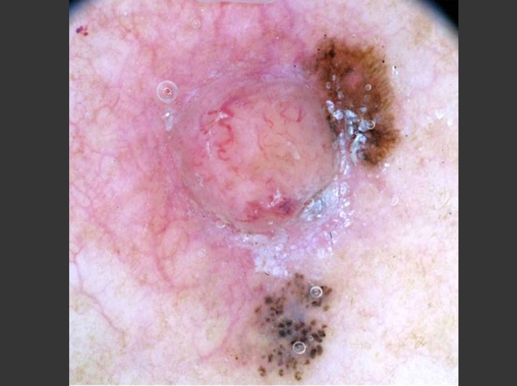
 Lesion on a 47 year old woman’s chest