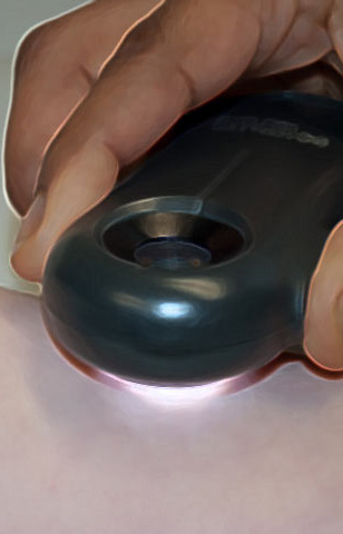
Virtual Dermatoscope
 Pigmented plaque, upper arm eldery lady, fair skin, severe sun-damage
 This plaque was found on a 60 year old man’s back
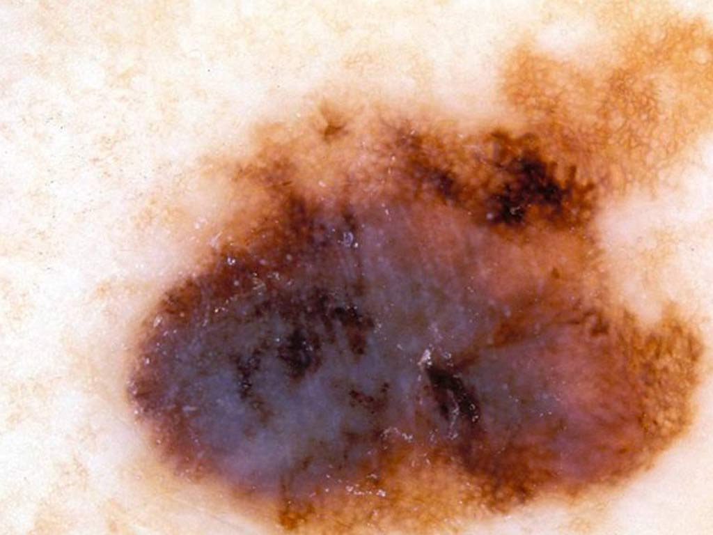
 Pigmented lesion on a 70 year old man’s back
 This is a plaque on a 45 year old man’s back
 A pigmented lesion on a 25 year old man’s foot
 A lesion on sundamaged skin on the neck of a 52 year old woman
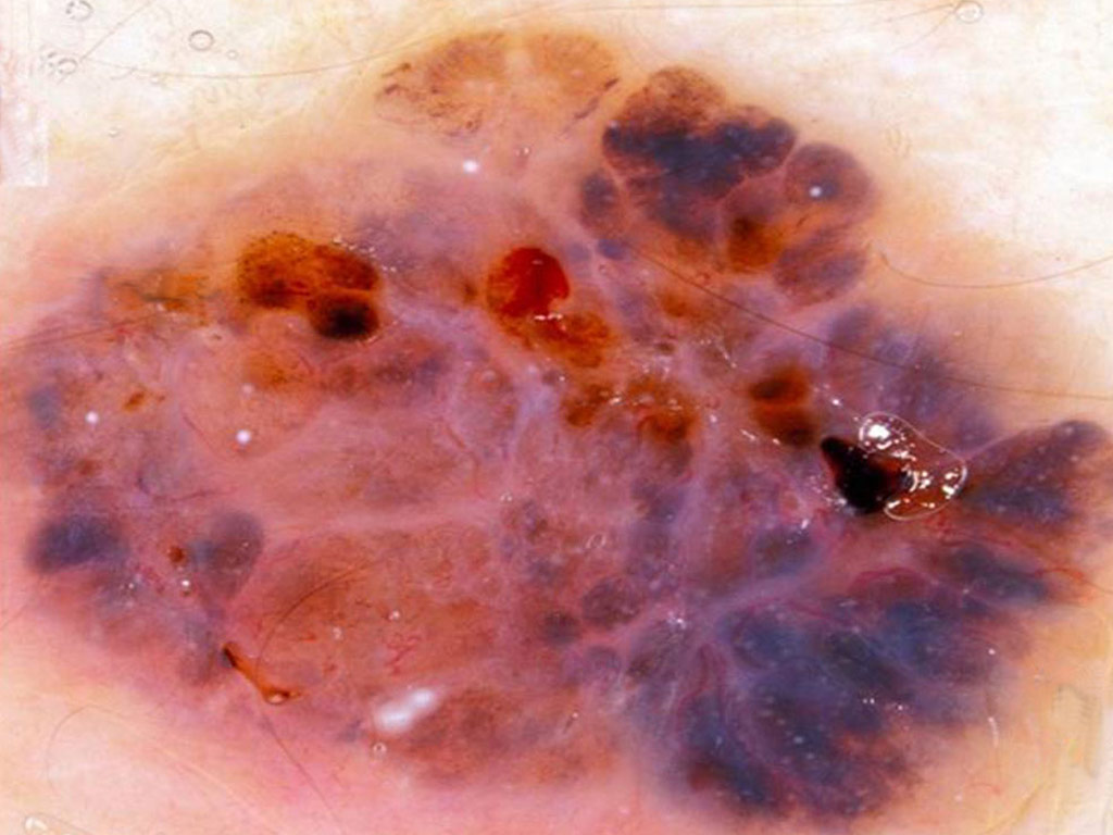
 A pigmented lesion on back of an elderly man
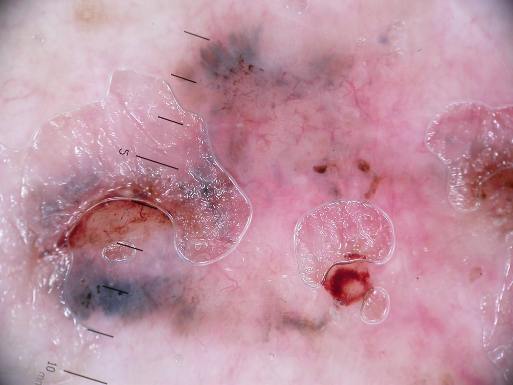
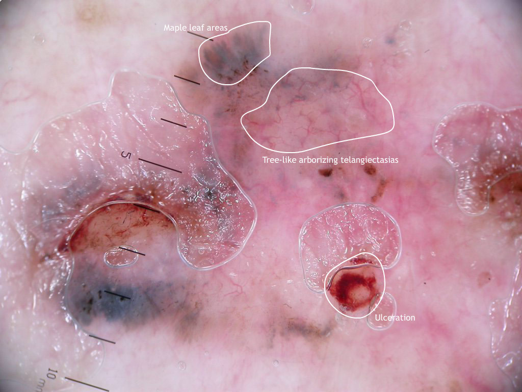
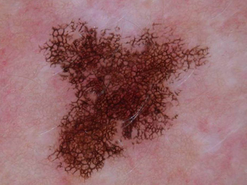
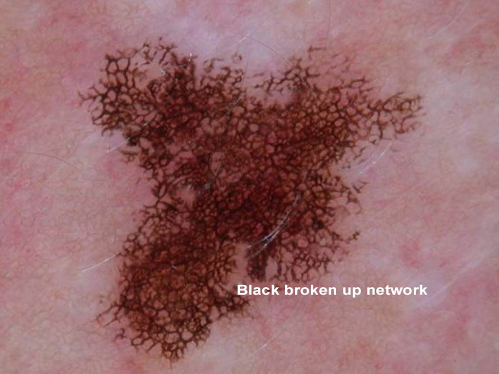
 This palpable pigmented lesion was found on a 42 year old man’s back
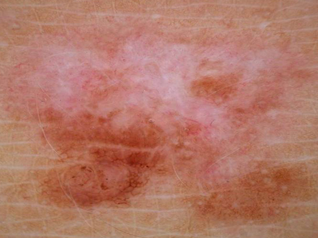
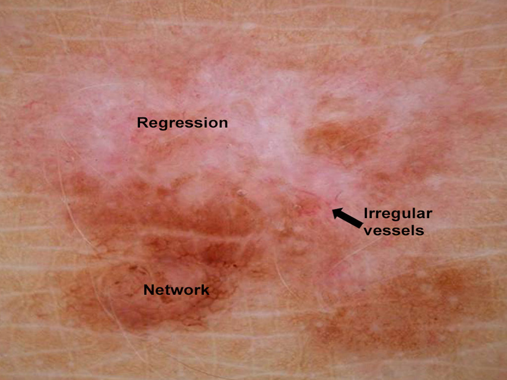
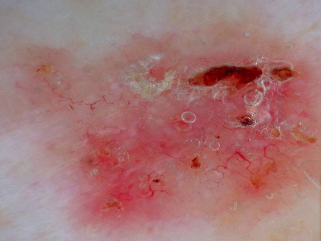
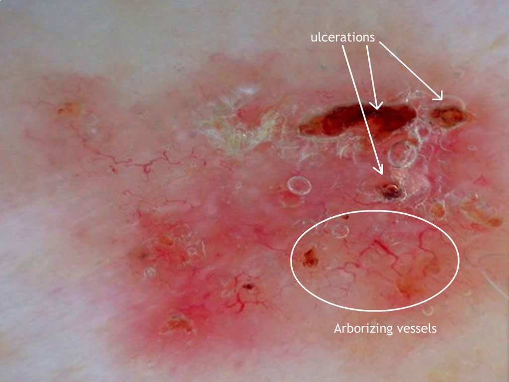
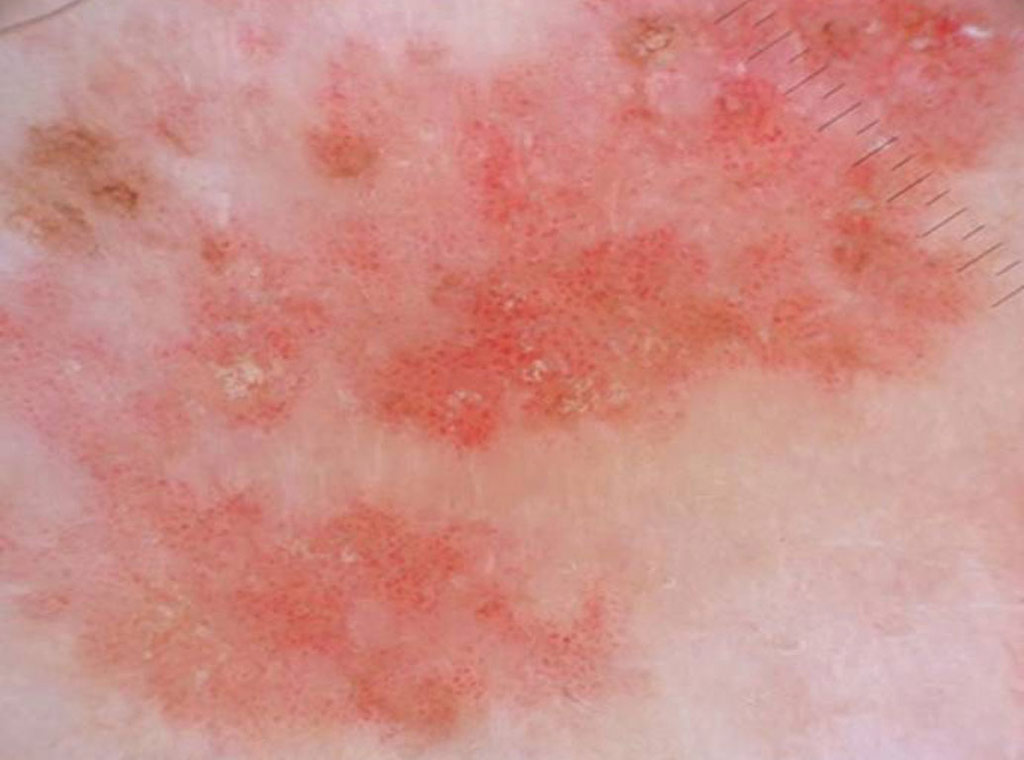
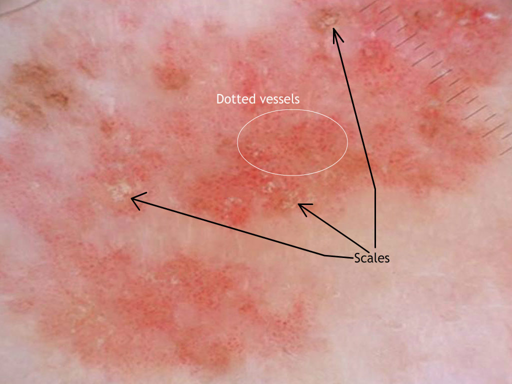
 This is a plaque on the shin of a 65 year old woman