Virtual Dermatoscope
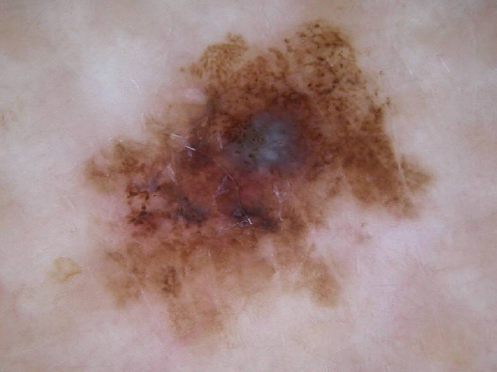
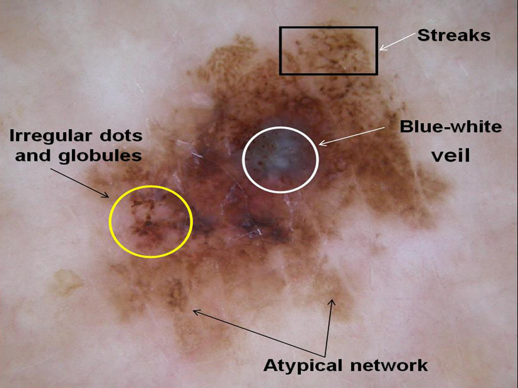
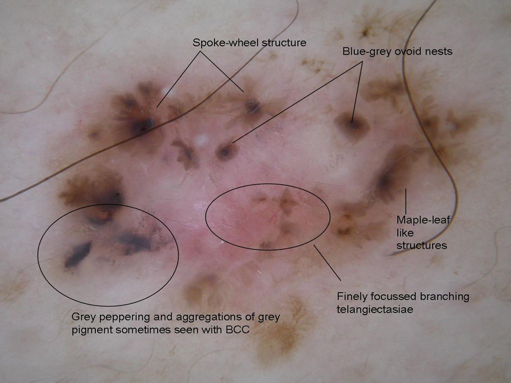
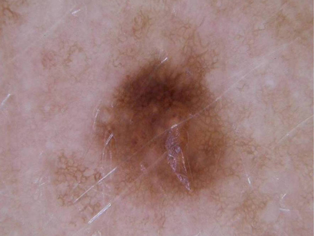
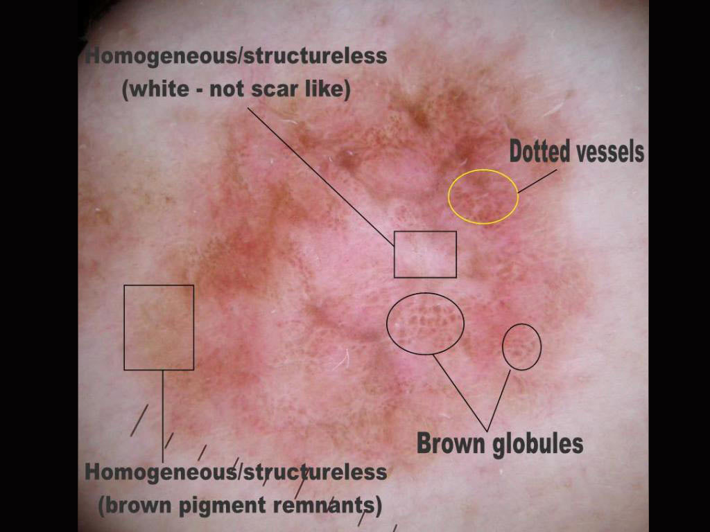
 A lesion on a 58 year old man’s thigh
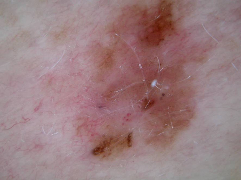
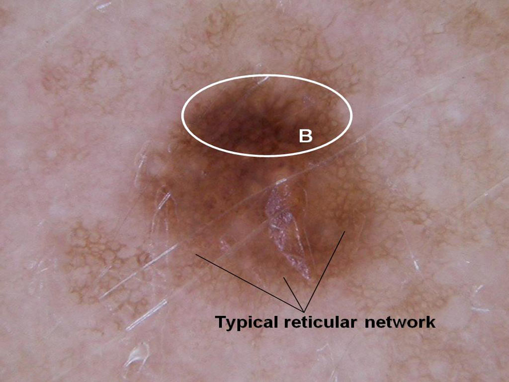
 A pigmented flat lesion on a 72 year old man’s neck
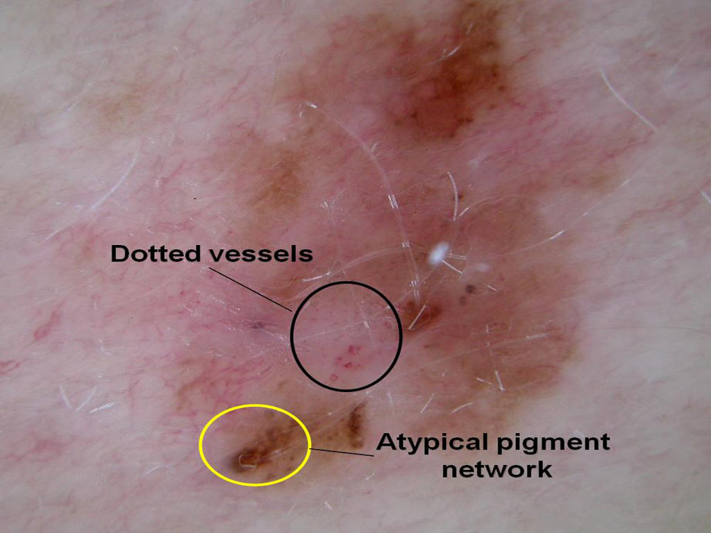
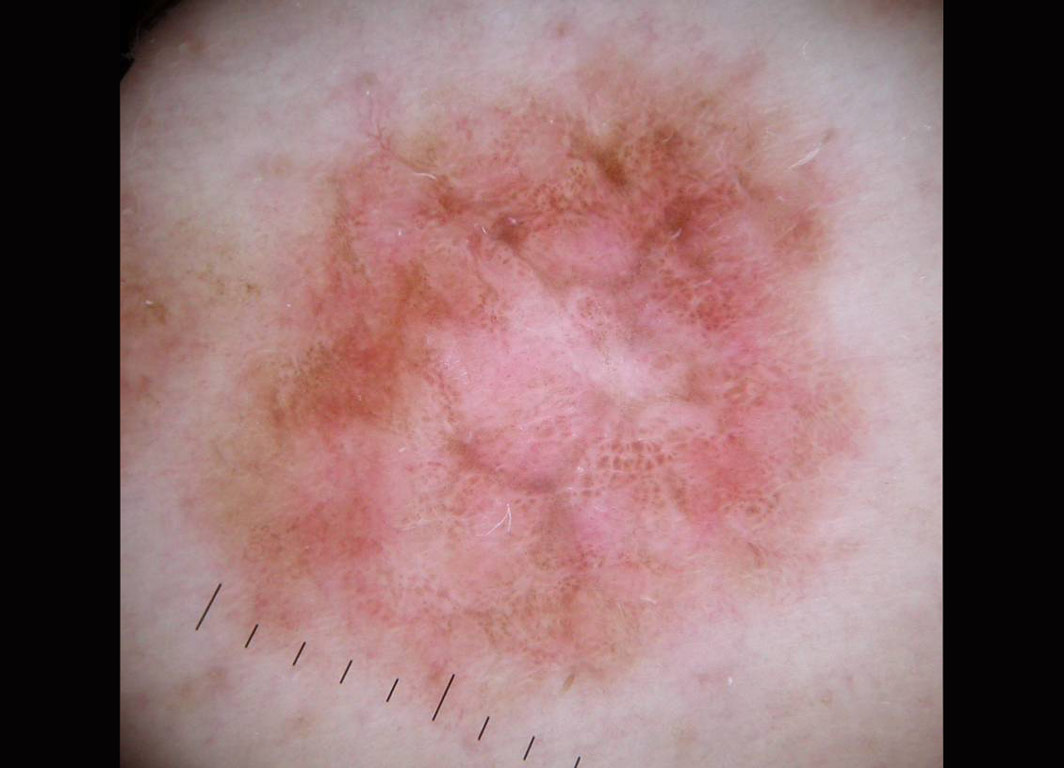
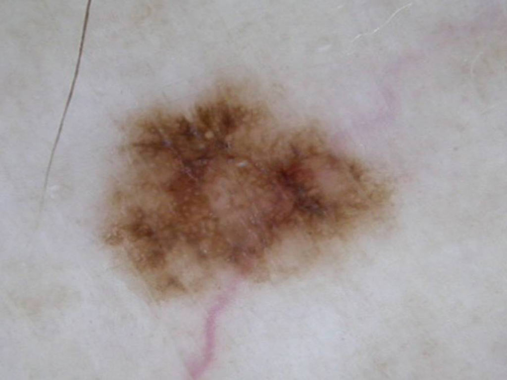
 A pigmented lesion on a 43 year old man’s back
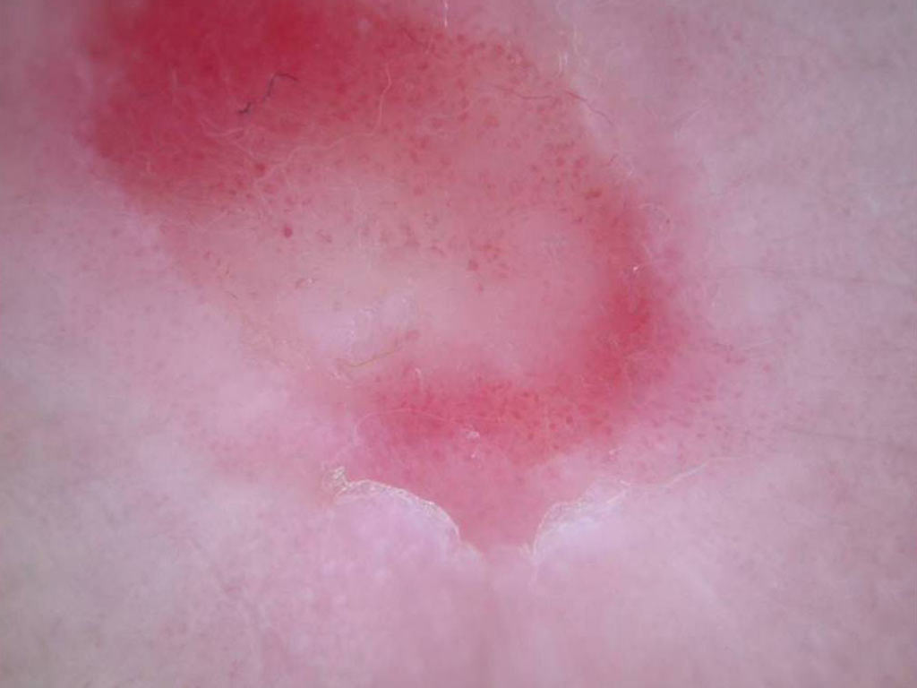
 A pigmented lesion on a 39 year old woman’s back
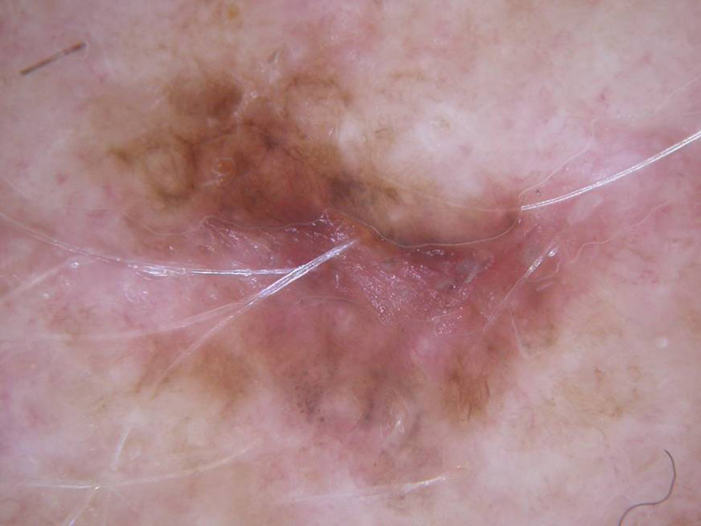
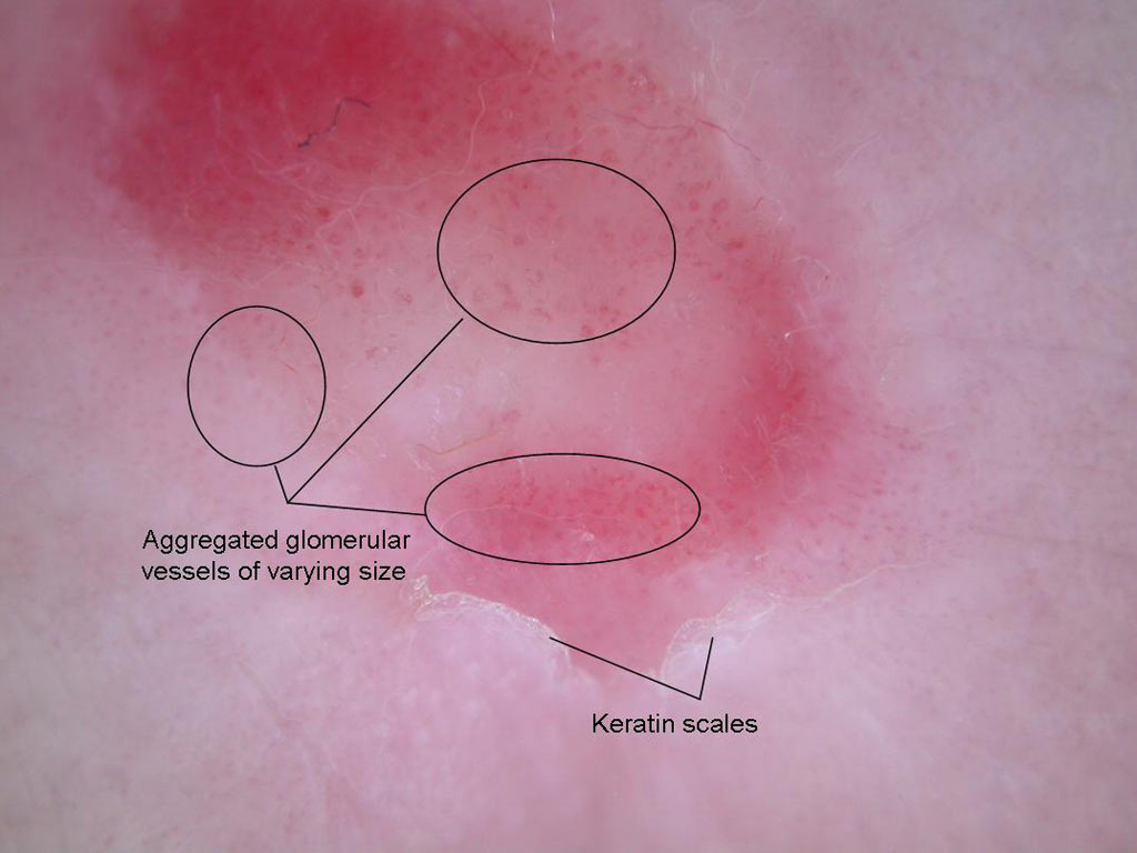
 An erythematous lesion on the dorsum of an elderly woman’s left hand
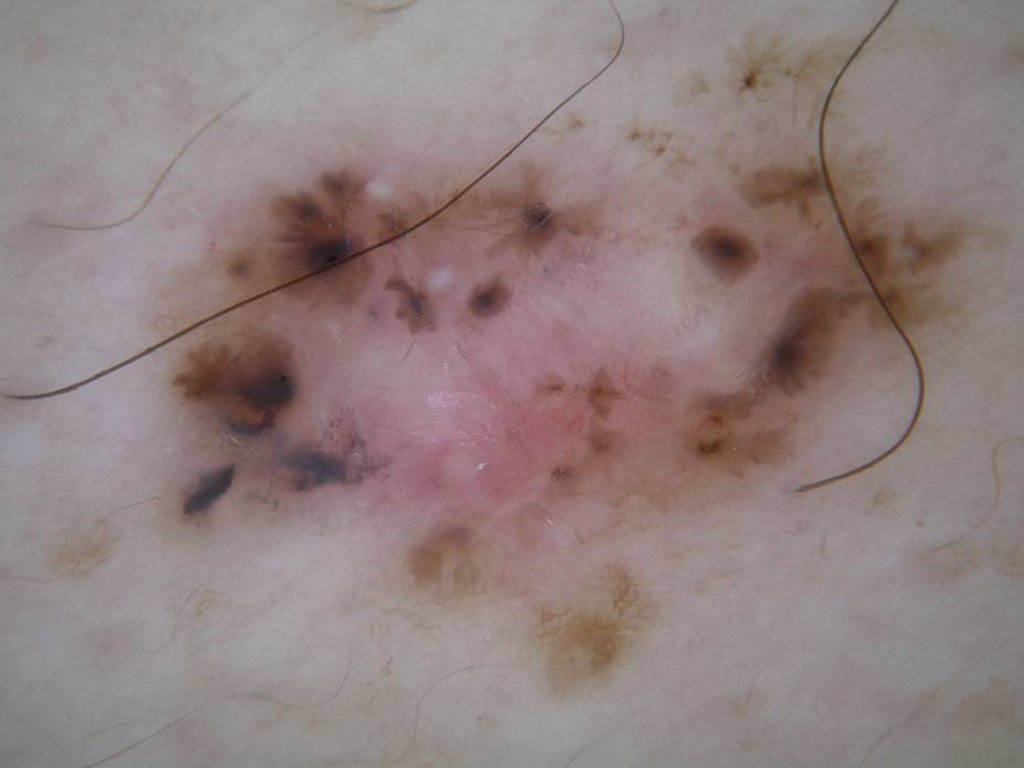
 A pigmented lesion on the back of a 32 year old woman
 A pigmented lesion on the back of a 45 year old man with multiple naevi
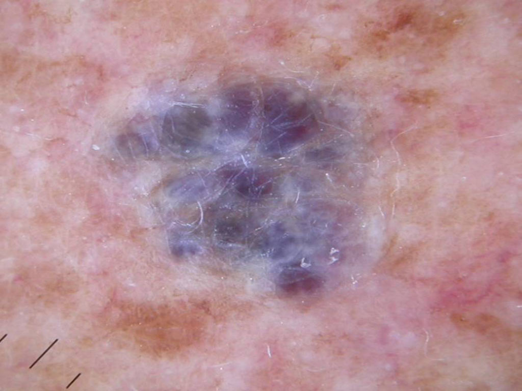
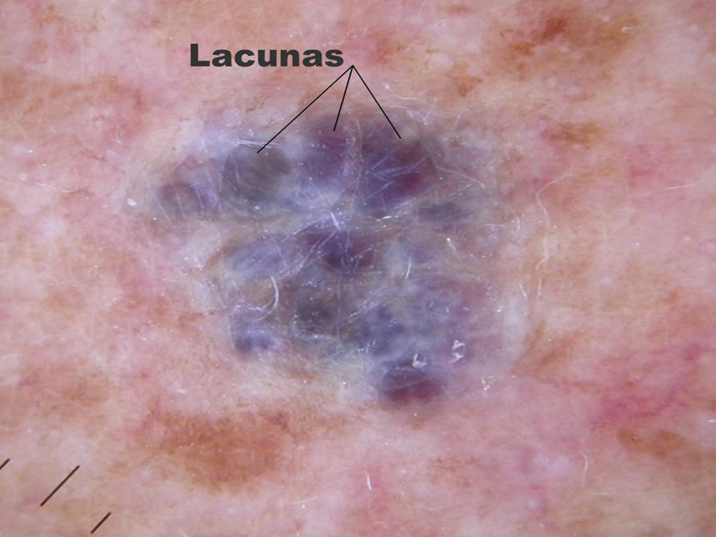
 A bluish lesion on the back of a 55 year old man
 A pigmented lesion on the back of a 52 year old man